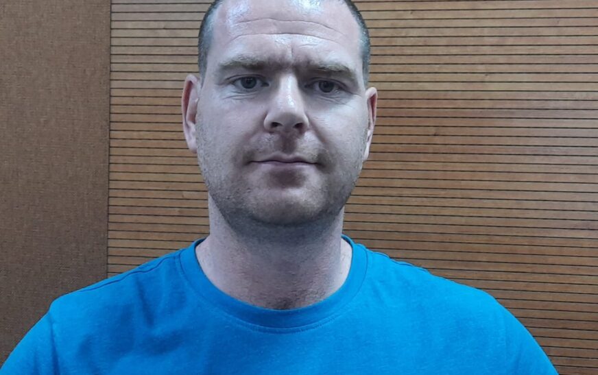
Dr. Ryan Beveridge, the Technical Services Coordinator at Ulster University‘s School of Computing, Engineering, and Intelligent Systems, is closely affiliated with the Northern Ireland Functional Brain Mapping Facility (NIFBM). His research focuses on brain-computer interfaces, neuroimaging, and electron photography.
In an exclusive interaction with The Interview World, Dr. Beveridge discusses cutting-edge technologies and methodologies used to explore brain pulses and muscular functions. He highlights the increasing adoption of these techniques in modern practice and emphasizes the current neuroimaging technologies employed in medical and research environments. Here are the key insights from his interview.
Q: What emerging technologies and methodologies are currently utilized to study and comprehend the dynamics of brain pulses and muscular functions?
A: In the dynamic realms of psychology and visual audio processing, the utilization of electroencephalography (EEG) signals has emerged as a pivotal avenue of exploration. These signals, emanating from the intricate interplay of neurons within the brain, offer a window into the depths of cognitive processes. Through sophisticated analysis techniques, researchers can dissect these signals, unraveling the mysteries of human cognition and behavior.
One remarkable application of EEG lies in the realm of brain-computer interfaces (BCIs), where the power of thought alone can commandeer computer functions. This revolutionary technology transcends the constraints of traditional input devices, offering a lifeline to individuals with muscular disabilities or impairments. By tapping into the neural symphony of the brain, BCIs empower users to interact with technology seamlessly, heralding a new era of accessibility and inclusivity.
Consider the profound implications of this technology. Imagine a world where typing emails, browsing the internet, or even playing video games can be accomplished effortlessly, solely through the power of one’s mind. Such advancements not only enhance quality of life for individuals with disabilities but also pave the way for groundbreaking innovations in human-computer interaction.
Yet, EEG is just one facet of the neuroimaging landscape. Enter Magnetoencephalography (MEG), a technique that harnesses the subtle magnetic fields generated by neuronal activity within the brain. This modality offers a complementary perspective, enriching our understanding of brain function and connectivity. By detecting these magnetic signatures, MEG provides insights into the intricate dance of neural circuits, unveiling the hidden dynamics of cognition.
MEG’s capabilities extend beyond basic neuroimaging; it holds promise as a diagnostic tool for neurological disorders such as epilepsy. Through meticulous analysis of MEG data, clinicians can discern aberrant neural patterns associated with epileptic seizures, enabling more accurate diagnosis and treatment planning. Moreover, MEG facilitates precise localization of epileptogenic zones, guiding surgical interventions with unparalleled accuracy and efficacy.
In the realm of medical rehabilitation, MEG emerges as a beacon of hope for individuals recovering from stroke-induced impairments. By deciphering neural signals originating from the motor cortex, therapists can tailor rehabilitation protocols to the specific needs of each patient. This personalized approach accelerates recovery, empowering individuals to regain lost motor function and reclaim their independence.
Indeed, the fusion of EEG and MEG technologies heralds a new frontier in neuroscience and rehabilitation. As our understanding of the brain’s intricate machinery deepens, so too do the possibilities for enhancing human health and well-being. From empowering individuals with disabilities to revolutionizing medical diagnostics and treatment, the potential of neuroimaging technologies knows no bounds. As we continue to unlock the secrets of the mind, we pave the way for a future where every individual can thrive, unencumbered by the limitations of their body or circumstance.
Q: What is the current prevalence or adoption rate of these techniques in contemporary practice?
A: Electroencephalography (EEG), a technique pioneered in 1921, has been a staple in neuroscience for over a century, serving diverse functions from medical diagnoses to facilitating Brain-Computer Interface (BCI) interactions. In contrast, the emergence of Magnetoencephalography (MEG) roughly two decades ago heralded a new era in neural signal analysis. MEG offers the promise of surpassing EEG’s capabilities in providing insights into brain activity, yet it grapples with challenges such as costliness and unwieldiness.
However, ongoing research endeavors are actively exploring MEG’s potential. Various studies have yielded promising results across a spectrum of neurological conditions. As researchers delve deeper into its applications, the horizon looks bright for MEG technology. With each new study published and breakthrough achieved, MEG inches closer to wider adoption and integration into clinical and research settings. The ongoing pursuit of understanding and harnessing neural signals through MEG promises to revolutionize our comprehension of the brain’s inner workings.
Q: What are the current neuroimaging technologies utilized in contemporary medical and research settings?
A: Several techniques are employed to measure brain activity, each offering unique insights into neural processes. Electroencephalography (EEG) involves the placement of electrodes on the scalp to capture electrical signals emitted by the brain. Renowned for its real-time data acquisition and high temporal resolution, EEG is instrumental in studying dynamic brain activities.
Similarly, Magnetoencephalography (MEG) measures the magnetic fields generated by neuronal activity, complementing EEG’s findings with its exceptional temporal and spatial resolution capabilities. This method enables researchers to delve deeper into the intricacies of brain functioning.
Functional Magnetic Resonance Imaging (FMRI) stands out for its ability to detect changes in blood flow associated with brain activity. It provides detailed spatial information, enhancing our understanding of cognitive processes and neurological disorders.
Lastly, Functional Near-Infrared Spectroscopy (fNIRS) offers a portable and non-invasive means of monitoring brain hemodynamics. By utilizing near-infrared light, fNIRS provides insights into cerebral blood flow and oxygenation levels, contributing to a holistic understanding of brain function.
Q: What are the distinctions between neuroimaging and MRI, and how do they relate to each other?
A: Certainly! Neuroimaging, a vast domain in neuroscience, surpasses the confines of MRI, a specific imaging modality. While MRI, or Magnetic Resonance Imaging, is a cornerstone of neuroimaging, the field encompasses an extensive array of techniques dedicated to exploring both the structure and function of the brain. Alongside MRI, other methodologies such as CT (Computed Tomography), PET (Positron Emission Tomography), EEG (Electroencephalography), and fMRI (functional Magnetic Resonance Imaging) play pivotal roles in unraveling the mysteries of the brain.
MRI stands out for its ability to generate detailed images of the brain’s anatomy using powerful magnetic fields and radio waves. However, neuroimaging extends beyond mere anatomical visualization; it ventures into the realm of understanding brain activity, connectivity, and metabolic processes. Techniques like fMRI and PET offer insights into brain function by measuring indicators such as blood flow and metabolic activity. In essence, neuroimaging serves as a multifaceted toolkit, facilitating a comprehensive exploration of the brain’s complexities. While MRI serves as a crucial cornerstone within this toolkit, its integration with other techniques allows researchers to delve deeper into understanding the intricate workings of the brain, advancing our knowledge of neuroscience and its applications in various fields, from medicine to psychology.


